Integrating Insights
Digital pathology sits at the heart of AI in healthcare. Bridging the gap between digital imaging data and its clinical interpretation - or ‘explainability’ - ensures AI tools remain accountable and trusted in medical practice.


Enhancing Outcomes
-
![Line drawing of an open coffee machine with visible internal components.]()
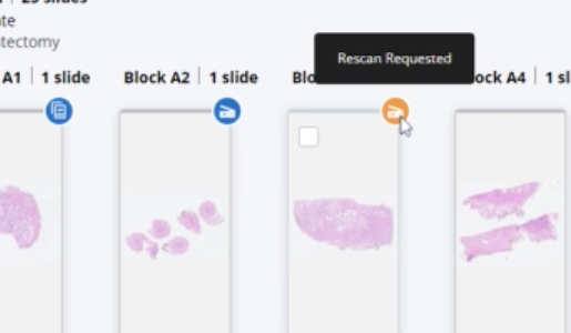
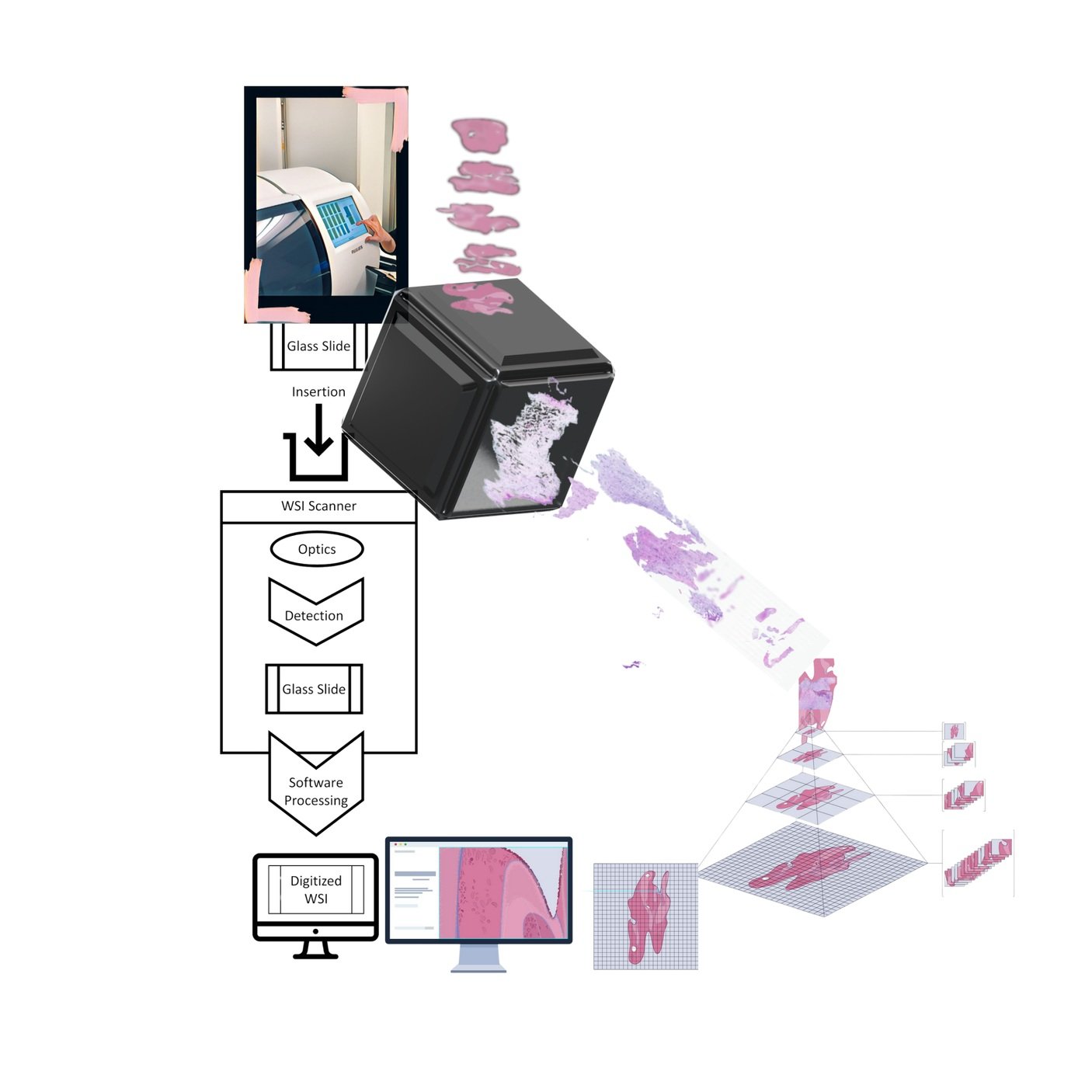
Laboratory professionals are navigating the transition from traditional microscopy to digital whole slide image (WSI) analyis.
-
![Illustration of a microscope examining a purple heart shape on a slide set on a stage against a light blue background.]()
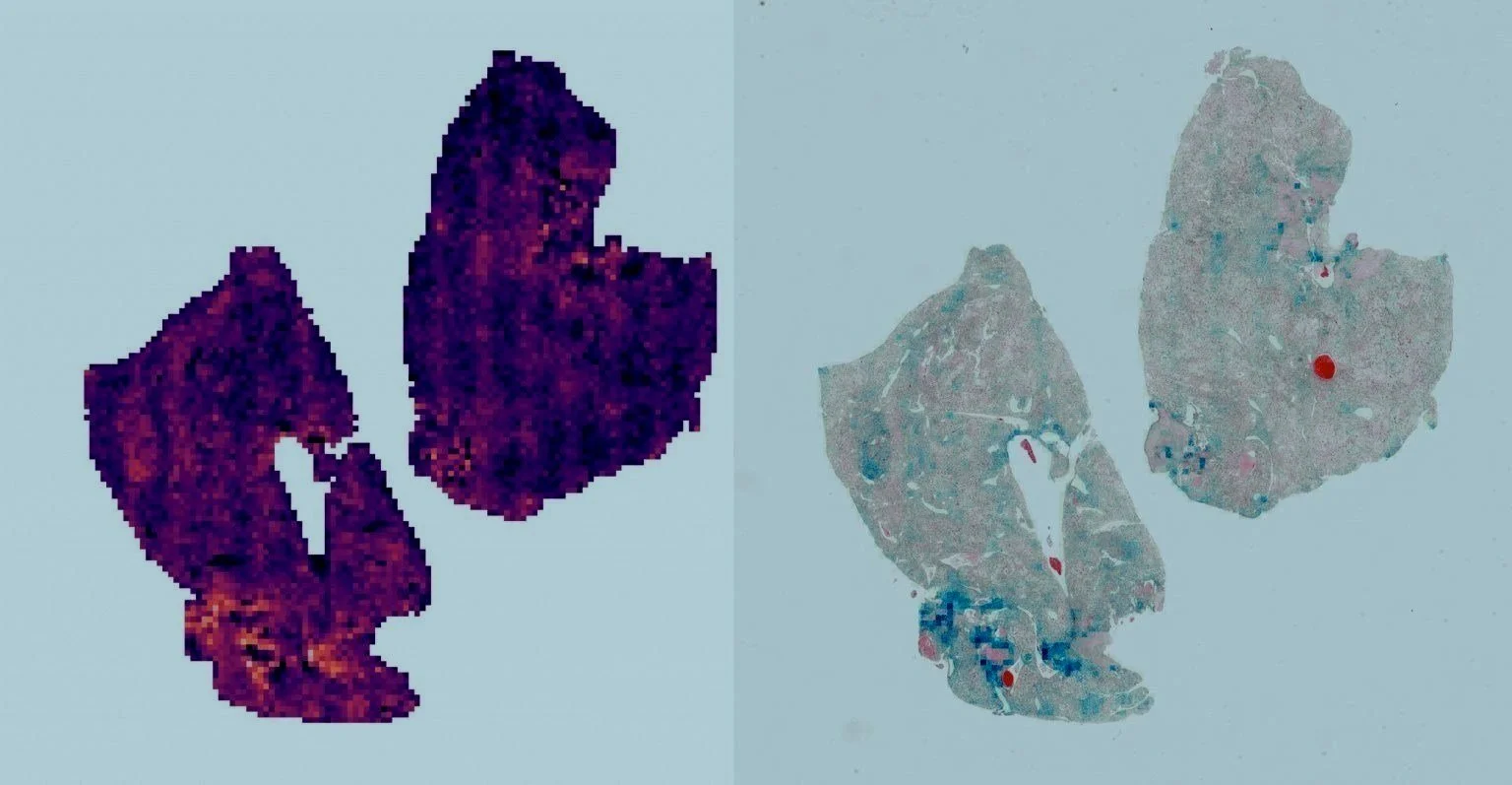
Digitization of patient samples is the foundation of explainability in AI.
-
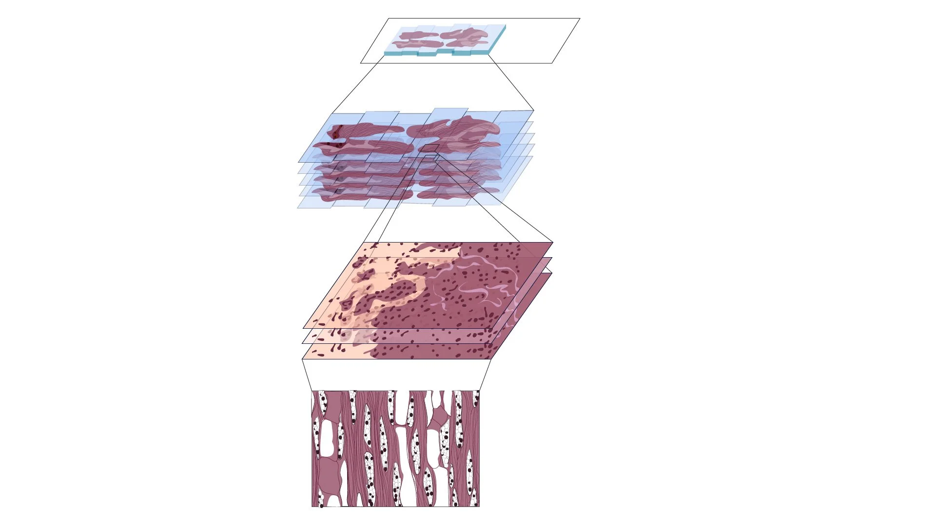
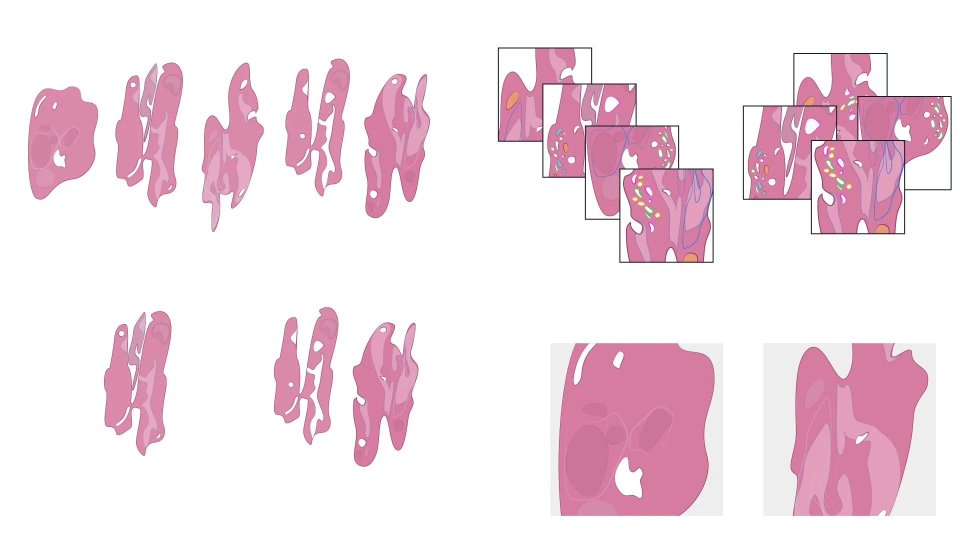
![Diagram of tissue layers in a cross-section, showing structure and details.]()
The initial step in ensuring transparency and interpretability in AI-driven diagnostics begins with patient images and their data.


-
![]()
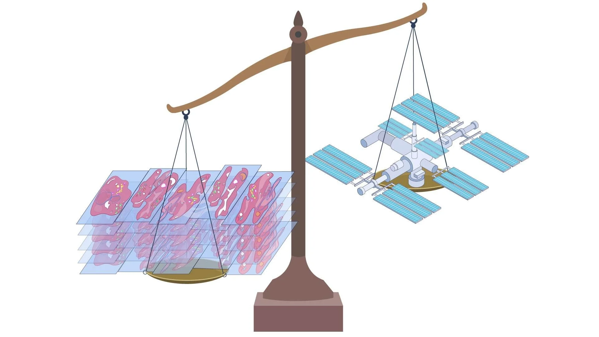
Digital pathology data is central to AI development, but whole slide images (WSI) pose significant storage and operational challenges. Data compression, cloud storage, and retrieval systems are just some of many important elements essential to this operation.
-
![Illustration of a balance scale with a stack of bricks on one side and a purple whale on the other, both appearing to weigh equally.]()
Average weight of a humpback whale = approximate volume of traditional Hollerith “punch” cards needed to store 1gb of WSI data
-
![Illustration of a balance scale with cells on one side and a space station on the other, suggesting a comparison or relationship between cell research and space exploration.]()
2x the weight of the International Space Station (ISS) = the average volume of glass slides handled yearly at major academic medical centers, large regional hospitals, and specialized reference laboratories serving multiple healthcare facilities
-
![Illustration of a balance scale with a brain on one side and a laptop displaying film icons on the other.]()
Data requirements for storing cumulative (new and archival) WSI data at a high-volume, fully digitized academic medical center (in PB) = approximately 20x the total storage capacity of typical major streaming service providers (Netflix, Amazon Prime Video, Hulu, etc.)
-
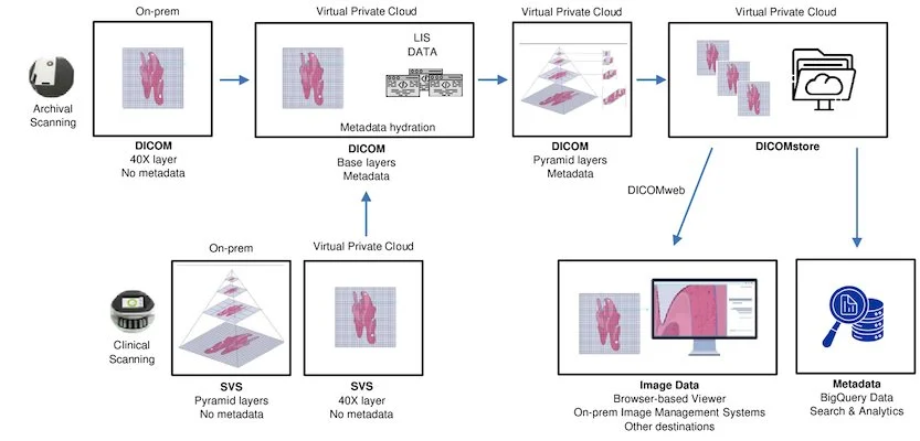
![Flowchart of a medical image processing system showing archival and clinical scanning. It depicts the transition of DICOM and SVS formats through on-premise and virtual private cloud, with metadata integration and storage options like DICOMstore, image viewers, and metadata analysis tools. Involves processes like 40X layering, base layering, pyramid layering, and LIS data utilization.]()
Successful laboratory digitization requires close collaboration between pathologists and IT specialists. By aligning technological infrastructure with clinical workflows, we ensure efficient, sustainable systems that enhance diagnostic capabilities.


Pathologists are stewards of laboratory data, establishing diagnostic ground truth for AI model validation. With 70% of medical decisions driven by laboratory diagnostics, pathologists' expertise forms the foundation of explainable AI. While current AI solutions focus on imaging, the future lies in integrating broader laboratory data.